
 |
 |
 |
 |
 |
 |
 |
 |
 |
 |
 |
 |
 |
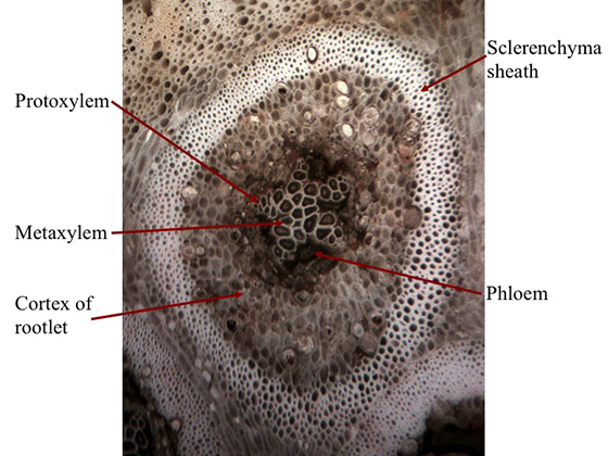 |
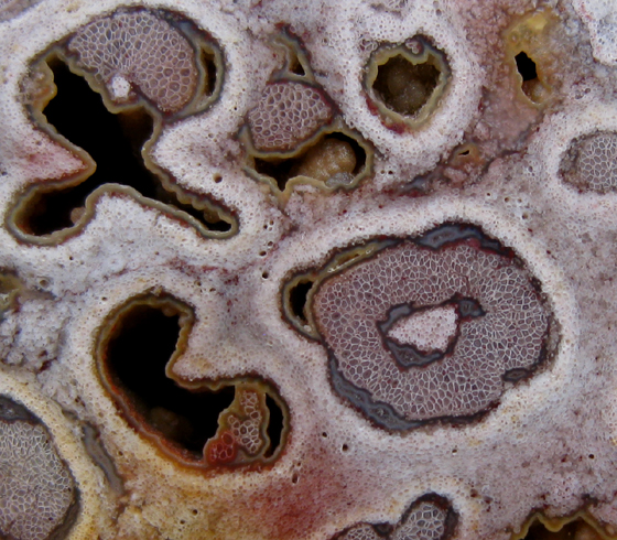
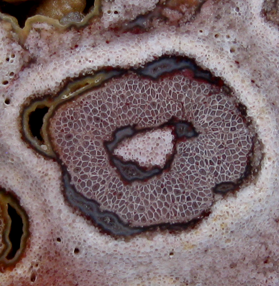
A cross-section of an aerial root found in the mantle of a tree fern reveals the central, star-shaped xylem. As the aerial root developed, primary xylem grew from five to nine different points. Metaxylem was then produced towards the center forming a star shape. Phloem tissue developed between the arms of the star. Cortical tissue and air spaces surround the xylem and phloem tissues. A sheath made of sclerenchyma cells enclosed the cortex. The root epidermis and cortical cells outside the rootlet proliferated to produce a dense mass of parenchyma tissue that held the root zone together. The aerial rootlet pictured above comes from a permian-aged Brazilian tree fern (Titea singularis). The picture was taken at 30x. Below one can see the specimen from which the micrograph was taken. To view a series of images that zoom in on the individual rootlet visit the Brazil Gallery (slide 1) in the Permian section of our website. Learn how to create a series of images that zoom in on a fossil specimen using a digital camera and microscope by reading Images of Cells Preserved in Stone. |
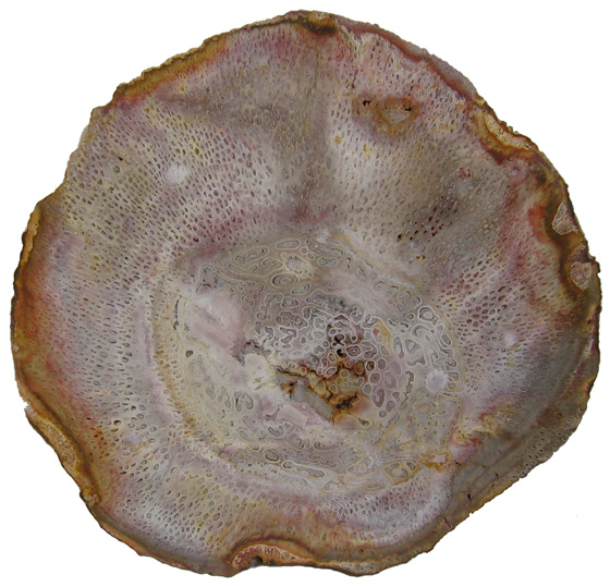 |
Tree
Fern Titea singularis Bieland, Maranhao Province, Brazil Pedra de Fogo Formation Permian 23 cm diameter |
| Close-up photographs of Titea singularis reveal plant tissues preserved in silica exhibiting beautiful pastel colors. The star-shaped xylem of the individual rootlets may conjure up images of snowflakes. |
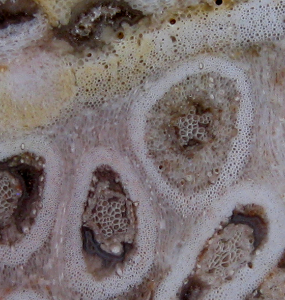 |
| In the image above one can see adventitious roots from the inner part of the mantle. Proliferating epidermis and cortical tissues appear to "flow" around the individual roots which they interconnect. |
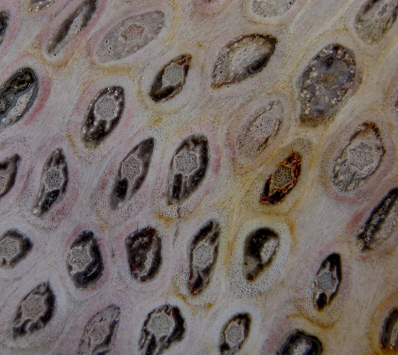 |
The
images below zoom in on vascular tissue found in the central vascular
cylinder of the specimen above. Phloem can be found on either side
of the xylem tissue. |
 |
 |