 |
 |
 |
 |
 |
 |
 |
 |
 |
 |
 |
 |
 |
Monocot Stems
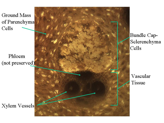 |
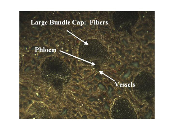
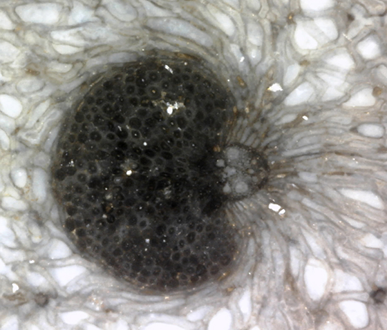
The vascular bundles (fibrovascular bundles) of palm fiber are composed of vascular tissue reinforced by a cap of thick-walled sclerenchyma cells. The large, water conducting xylem vessels are accompanied by much smaller water conducting tracheids. Air spaces are sometimes found in the vascular tissue area as well. The food conducting phloem tissue is found between the vessels and the bundle cap. Sieve tube members associated with companion cells make up the phloem tissue. Sieve tube members transport the products of photosynthesis through the plant. Companion cells are parenchyma cells that develop from the same mother cell as the sieve tube member. The companion cell helps transport materials in and out of the sieve tube member. A space occupies the area in which the phloem would have been situated. Phloem is only rarely preserved. Visit our Texas Gallery in the Oligocene section of our website to see vascular bundles with preserved phloem. The bundles caps of palm fiber are embedded in a ground mass of parenchyma cells. The vascular bundle pictured above measures 1,400 micrometers tall and a little under 900 micrometers at its widest point. The vessels ("eyes") measure a little over 200 micrometers in diameter. Visit the Louisiana Gallery (slide 20) in the Oligocene section of our website to see a sequence of photos that zoom in to the photo above. |
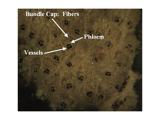 |
| The individual vascular bundles can be seen in this fossil palm (genus Palmoxylon) specimen from Wyoming. Each vascular bundle is surrounded by numerous fibers, which thicken into a cap shape on one end. The fibers provide structural support. The empty spaces represent vessels for water conduction and sometimes air spaces. The phloem tissue would be found between the vessels and the bundle cap. This image was taken at 40x. |
 |
| The fossil Palmoxylon pictured above comes from the same location in Wyoming as the previous specimen. Note this palm has much larger bundle caps in comparison to the vascular tissues. |
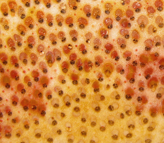 |
The colorful vascular bundles in this trunk of fossil Palmoxylon from Louisiana exhibit great cellular detail. In some species of palm one can observe fibrous bundles, which are made of the same cells making up the bundle caps in the fibrovascular bundles. Fibrous bundles are made of fibers and appear as red and orange dots in the specimen above. Note the Wyoming specimens above lack the fibrous bundles. The next image zooms in on a fibrovascular bundle and fibrous bundle at 150x with a Dino-Lite AD7013 MT 5.0 MP. The image was resized in Adobe Photoshop CS6. |
 |
The Palmoxylon trunk used to take the images above is pictured below. |
 |
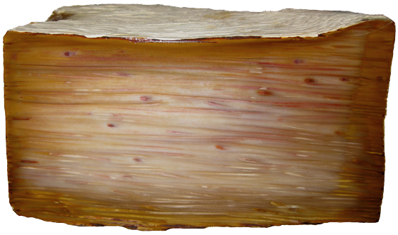
A longitudinal cut Palmoxylon specimen from Louisiana reveals the rod-like structures of the vascular bundles. |
|
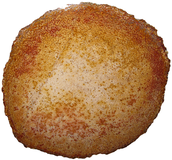
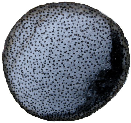
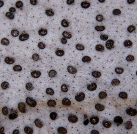
This Palmoxylon specimen from Texas represents a transverse cut (cross-section). When viewed in cross-section the vascular bundles give the palm fiber a spotted appearance. |
 |
|
 |
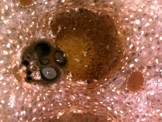
The close-up below shows a vascular bundle at 150x and was taken with a Dino-Lite AD7013 MT 5.0 MP. The image was resized in Adobe Photoshop CS6. |
 |
 Oligocene OligoceneCatahoula Formation Fayette County, TX |
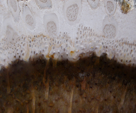
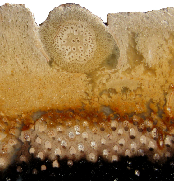
The images above and below illustrate the transition zone between Palmoxylon trunks and their adventitious roots (Rhizopalmoxylon). In the image above individual rootlets measure 2 to 3 mm in diameter. The adventitious root below measures 4 mm in diameter. |
 Palmoxylon with Rhizopalmoxylon Palmoxylon with RhizopalmoxylonCenozoic; Tertiary East of Barisan Mountain Range Sumatra, Indonesia |